
Structure of a Neuron: 8 Parts, What Each Does, and the One Most Students Overlook
Reach for your coffee mug, and before your fingers close around the handle, a chain of cells has already carried a message from your brain to your hand. Each of those cells, a neuron, works like a relay runner: one end catches signals, a middle region decides whether to pass them on, and the far end hands them off. The structure of a neuron isn’t random. Every part, from the branching dendrites to the tiny gaps at the axon’s tip, exists to move a signal fast and without errors.
Key Takeaways
- A neuron has three jobs: receiving, deciding and sending. Each of its eight main parts serves one of them, which makes the whole diagram far easier to remember.
- The part that decides whether a neuron fires is one that many diagrams barely label, yet it controls everything that happens next.
- A neuron can’t fire harder when a stimulus gets stronger. Your brain uses a different trick to tell a light tap from a hard shove.
- Neurons never actually touch. The tiny gap between them is where caffeine, antidepressants and many other drugs do their work.
- Learning physically changes the connections between neurons, which is why practice strengthens skills and memories over time.
Why the Structure of a Neuron Matters
Your brain contains roughly 86 billion neurons, along with a similar number of non-neuronal cells (Azevedo et al., 2009).
The structure of a neuron makes the most sense when you treat it as a design solution. A neuron needs to collect information, decide whether it matters, and pass it on quickly. Each part handles one piece of that job. Neurons then link into larger circuits, including the crossed wiring that lets each brain hemisphere control the opposite side of your body.
The 8 Parts of a Neuron
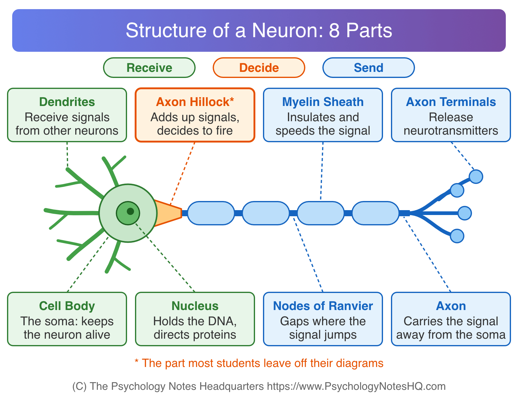
Textbooks label the structure of a neuron in slightly different ways, but most include the eight parts below. As you read, sort each one into a job: receiving, deciding or sending.
Dendrites: The Receivers
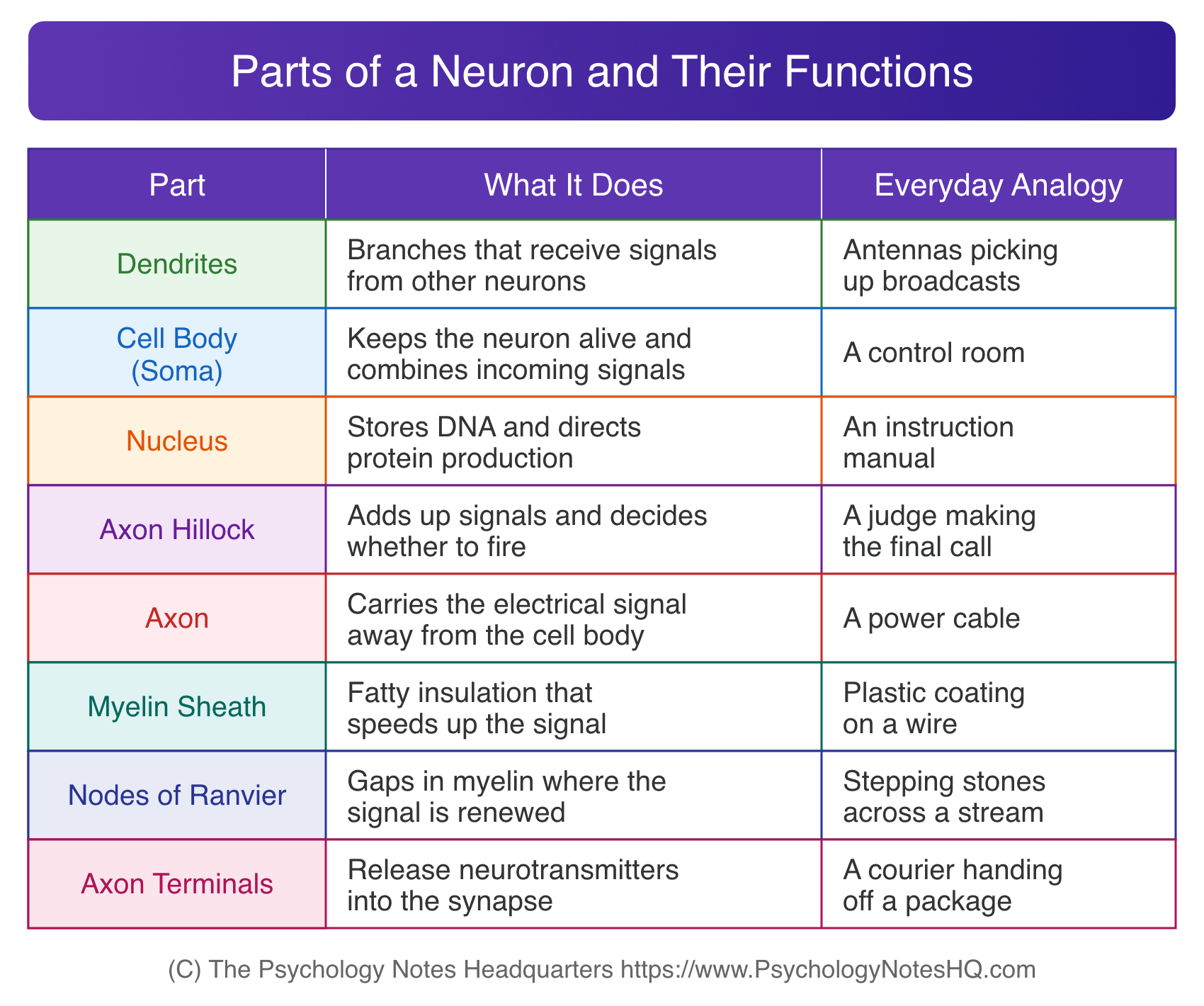
Dendrites are the branching extensions that grow out of the cell body. They serve as input sites, picking up signals from neighboring neurons (Spielman et al., 2020).
The branching matters. A neuron with more dendrite branches can collect input from more cells at once. Many dendrites are also covered in tiny bumps called dendritic spines, which become important later when we look at learning.
The Cell Body (Soma): The Life Support
The cell body, or soma, is the neuron’s main hub. It contains the machinery that keeps the cell alive, including structures that produce energy and build proteins.
It also plays a role in signaling. Messages arriving from the dendrites spread across the soma, where they combine. Some push the neuron toward firing, and others hold it back. The overall balance then moves on to the next part of the cell.
The Nucleus: The Instruction Manual
The nucleus sits inside the soma and holds the neuron’s genetic material (Spielman et al., 2020). It directs which proteins the cell makes, including the receptors and channels the neuron needs to send and receive signals.
Students sometimes label the nucleus as the neuron’s “decision maker.” That’s a mistake. The nucleus handles long-term maintenance, while the moment-to-moment decision about firing happens somewhere else.
The Axon Hillock: The Decision Point
Here’s the part many diagrams skip. The axon hillock is the cone-shaped region where the axon joins the cell body. It works like a gatekeeper, adding up all the signals that reach it.
If the combined input crosses a set threshold, the neuron launches an action potential, the electrical signal that travels down the axon. If the input falls short, nothing is sent. That makes this small region the true decision point of the neuron.
The Axon: The Cable
The axon is a long, thin fiber that carries the signal away from the cell body. Most neurons have a single axon, though it often splits into several branches near its end.
Axon length varies enormously, from a fraction of an inch to several feet (Spielman et al., 2020). The longest ones belong to neurons that run from the lower spinal cord all the way down to the foot.
The Myelin Sheath: The Insulation
Many axons are wrapped in myelin, a fatty coating made by glial cells. In the brain and spinal cord, cells called oligodendrocytes produce it, while Schwann cells do the job in the rest of the body (Purves et al., 2001).
Myelin works like the plastic coating on a wire, and the speed difference is dramatic. Unmyelinated axons conduct signals at about 0.5 to 10 meters per second, while myelinated axons can reach up to 150 meters per second (Purves et al., 2001).
Nodes of Ranvier: The Stepping Stones
Myelin doesn’t cover the axon in one continuous tube. It comes in segments, separated by small gaps called the nodes of Ranvier (Spielman et al., 2020).
These gaps are where the signal gets renewed. Instead of moving smoothly along every bit of the axon, the electrical signal leaps from node to node, a process called saltatory conduction. That jumping is a big part of why myelinated neurons are so fast.
Axon Terminals: The Senders
At the far end, the axon branches into small knobs called axon terminals, also known as terminal buttons. Each terminal holds synaptic vesicles, tiny sacs filled with chemical messengers called neurotransmitters (Spielman et al., 2020).
When the electrical signal arrives, the terminals release these chemicals. This is the handoff point, where the neuron’s electrical message becomes a chemical one.
How Neuron Structure Powers the Action Potential
Almost every part of a neuron exists to create, carry or pass on one signal: the action potential. Once you understand how that signal works, the shape of the axon hillock, the myelin and the nodes suddenly makes sense.
Resting Potential: A Charged Battery
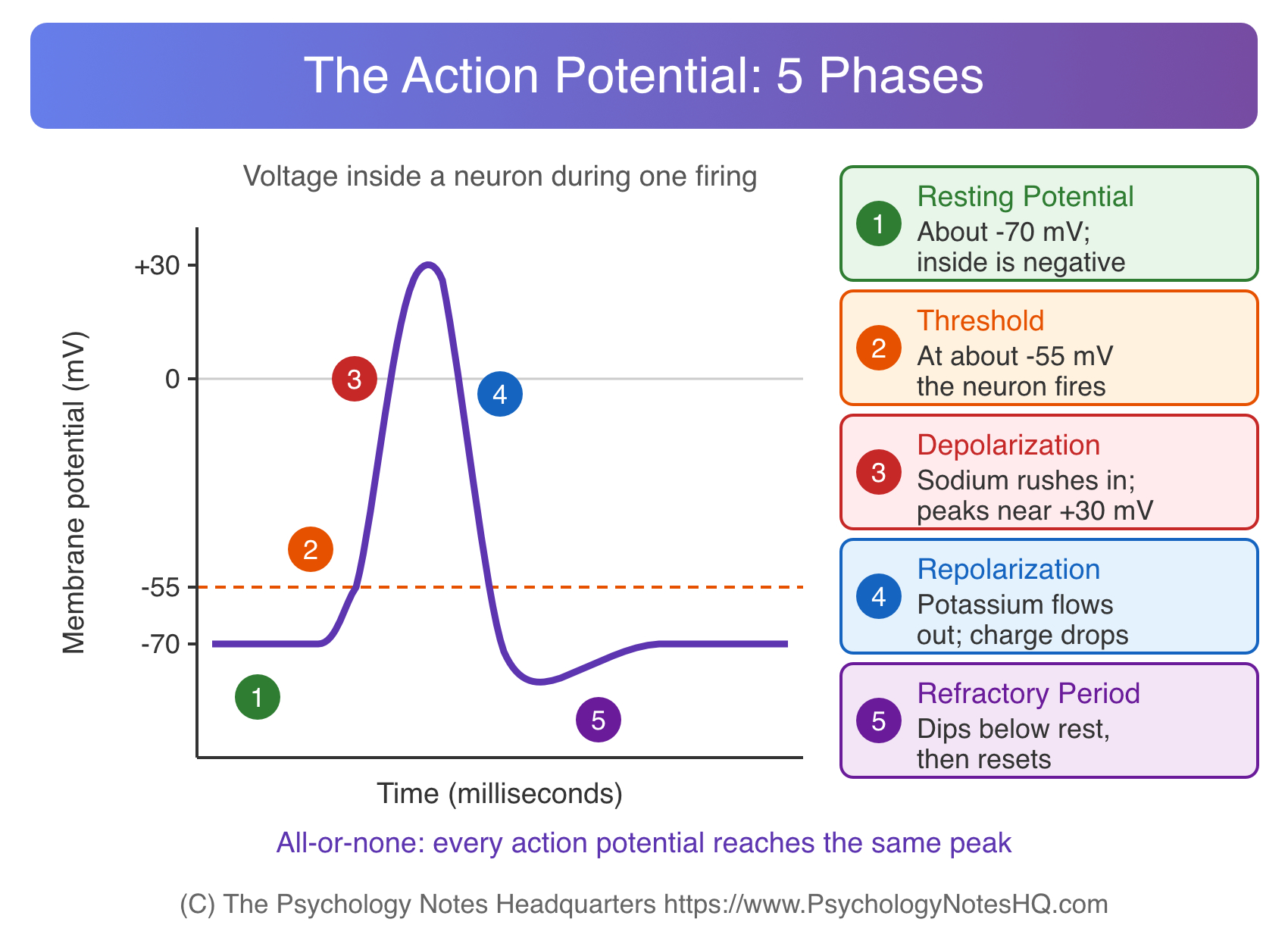
Between signals, a neuron sits at its resting potential, about -70 millivolts (Spielman et al., 2020). The negative value means the inside of the cell carries a more negative charge than the outside.
This difference comes from ions. Sodium is more concentrated outside the cell and potassium inside, and a sodium-potassium pump maintains the imbalance by moving three sodium ions out for every two potassium ions in. The neuron is loaded and ready, like a stretched rubber band.
Threshold and the All-or-None Principle
When incoming signals open gates in the membrane, sodium flows in and the inside becomes less negative. If the charge reaches about -55 millivolts, the threshold, the neuron fires (Spielman et al., 2020).
This follows the all-or-none principle. A neuron either fires completely or not at all. There’s no half-strength signal, and once it starts, it can’t be called back. The signal also regenerates at full strength along the axon, so it doesn’t fade on long trips.
How the Brain Codes a Stronger Stimulus
If every signal is the same size, how do you tell a light touch from a hard pinch? The answer lies in timing and numbers.
A stronger stimulus makes a neuron fire more often, packing more signals into each second. It also activates more neurons at the same time. Your brain reads intensity from these patterns rather than from the size of any single signal. This is one of the most commonly missed exam points about neurons.
The Refractory Period: Why Signals Travel One Way
At the peak, around +30 millivolts, sodium gates close and potassium flows out, pulling the charge back down. The neuron briefly dips to about -80 millivolts before settling at rest (Spielman et al., 2020).
During this reset, called the refractory period, the patch of axon that just fired can’t fire again right away. That keeps the signal moving forward. Hodgkin and Huxley (1952) mapped these sodium and potassium currents using the giant axon of a squid, work that later earned them a Nobel Prize.
The Synapse: How Neurons Communicate
Neurons never physically touch. Between the axon terminal of one neuron and the dendrite of the next sits a tiny space called the synaptic cleft (Spielman et al., 2020). The full junction, including both sides and the gap, is the synapse.
This gap is where electrical signals turn into chemical ones. Understanding how neurons communicate across it explains everything from reflexes to the effects of everyday drugs.
Synaptic Transmission in Four Steps
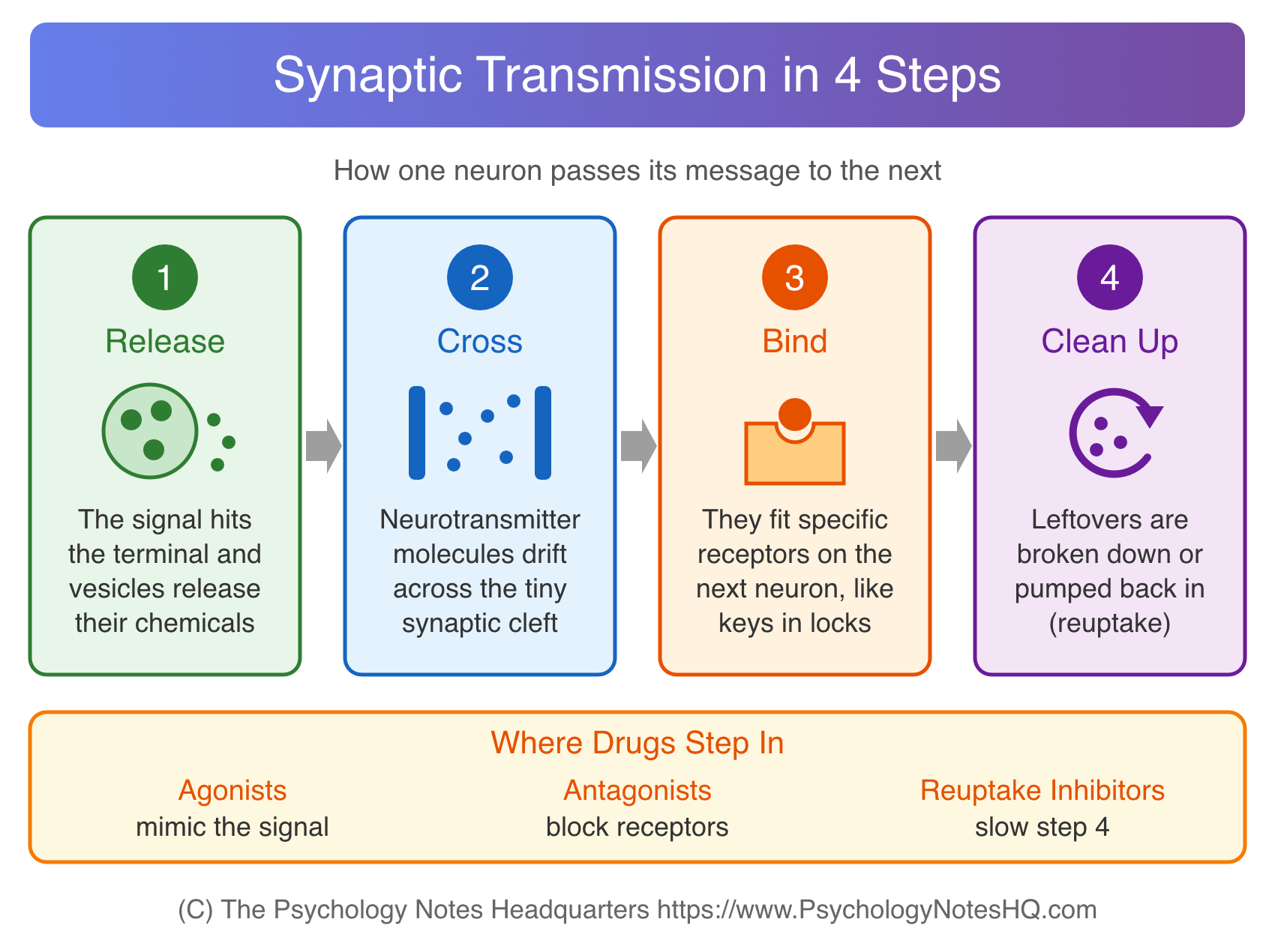
Synaptic transmission follows a predictable sequence (Spielman et al., 2020):
- Release: The signal reaches the axon terminal, and vesicles release neurotransmitters into the cleft.
- Cross: The molecules drift across the gap.
- Bind: They attach to receptors on the next neuron, each neurotransmitter fitting specific receptors like a key in a lock.
- Clean up: Leftover molecules are broken down or pumped back into the sending neuron through reuptake.
Excitatory and Inhibitory Signals
Not every message says “fire.” Excitatory signals push the receiving neuron closer to its threshold, while inhibitory signals push it further away. Glutamate is the brain’s main excitatory neurotransmitter, and GABA is its main inhibitory one.
A single neuron can receive thousands of these inputs, and the axon hillock tallies them all. Neurotransmitters also shape learning. Dopamine, for example, plays a central role in reward, linking synaptic activity to the way consequences shape behavior in operant conditioning.
How Drugs Act on the Synapse
Because so much happens at the synapse, it’s the main target for drugs that change mood, alertness and behavior. Most of them work in one of two ways. They either act on receptors, or they change how long neurotransmitters stay in the cleft.
Agonists and Antagonists
Agonists and antagonists both work at receptor sites, but with opposite effects. An agonist mimics a neurotransmitter and activates its receptor, while an antagonist blocks the receptor and stops the normal signal (Spielman et al., 2020).
Your morning coffee is a good example. Caffeine acts as an antagonist at adenosine receptors (Fredholm et al., 1999). Adenosine normally dampens neural activity, so blocking it leaves you more alert. That lift connects to arousal theory and why a little stimulation can boost performance.
Reuptake Inhibitors
Reuptake inhibitors take a different route. Instead of acting on receptors, they slow the cleanup step, so neurotransmitters stay active in the synaptic cleft longer (Spielman et al., 2020).
Selective serotonin reuptake inhibitors (SSRIs), a common class of antidepressants, work this way with serotonin. People usually take them for several weeks before noticing improvement, a delay researchers are still working to fully explain.
Three Types of Neurons, Three Different Shapes
Neurons share the same basic parts, but their shapes vary with their jobs.
Sensory neurons carry information from your senses inward. Many have a unipolar shape, with one process that splits in two. Motor neurons carry commands to muscles and glands, and most are multipolar, with many dendrites and one axon. Interneurons link neurons inside the brain and spinal cord. Bipolar neurons, with one dendrite and one axon, appear in the retina.
That coffee mug moment uses all three: sensory neurons report, interneurons process, motor neurons move your hand.
Glial Cells: The Support Crew
Neurons get most of the attention, but they don’t work alone. Glial cells provide the scaffolding of the nervous system, insulate axons, transport nutrients and waste, and help with immune defense (Spielman et al., 2020).
You may have read that glia outnumber neurons ten to one. That old figure doesn’t hold up. Azevedo et al. (2009) counted about 86 billion neurons and about 85 billion non-neuronal cells in the adult human brain, a ratio close to one to one.
How Neuron Structure Changes When You Learn
The structure of a neuron isn’t fixed. Connections between neurons strengthen, weaken and even grow in response to experience. This flexibility is the physical basis of learning.
Neurons That Fire Together Wire Together
Psychologist Donald Hebb (1949) proposed that when one neuron repeatedly helps fire another, the connection between them becomes more efficient. The catchy summary “neurons that fire together wire together” came later, but the core idea is his.
Hebb’s rule fits neatly with learning by association. When a bell keeps sounding right before food, the neurons processing each event fire together, and their link grows stronger. That’s the same logic behind how Pavlov’s dogs learned to salivate at a bell.
Long-Term Potentiation
Hebb’s idea got strong experimental support decades later. Bliss and Lømo (1973) stimulated a pathway in the rabbit hippocampus with brief trains of repeated pulses and found that the synaptic response stayed stronger for hours afterward.
This effect, now called long-term potentiation (LTP), is widely studied as a cellular mechanism of memory. It helps explain how experiences get stored as lasting episodic and semantic memories.
Common Mistakes When Labeling a Neuron
A few errors show up again and again on psychology exams:
- Leaving out the axon hillock, or labeling the nucleus as the part that decides whether to fire.
- Treating the synapse as part of one neuron. It’s the junction between two cells.
- Drawing myelin as one continuous tube. It’s segmented, with nodes of Ranvier in between.
- Saying a stronger stimulus produces a bigger action potential. It produces more frequent signals and recruits more neurons.
The Big Picture
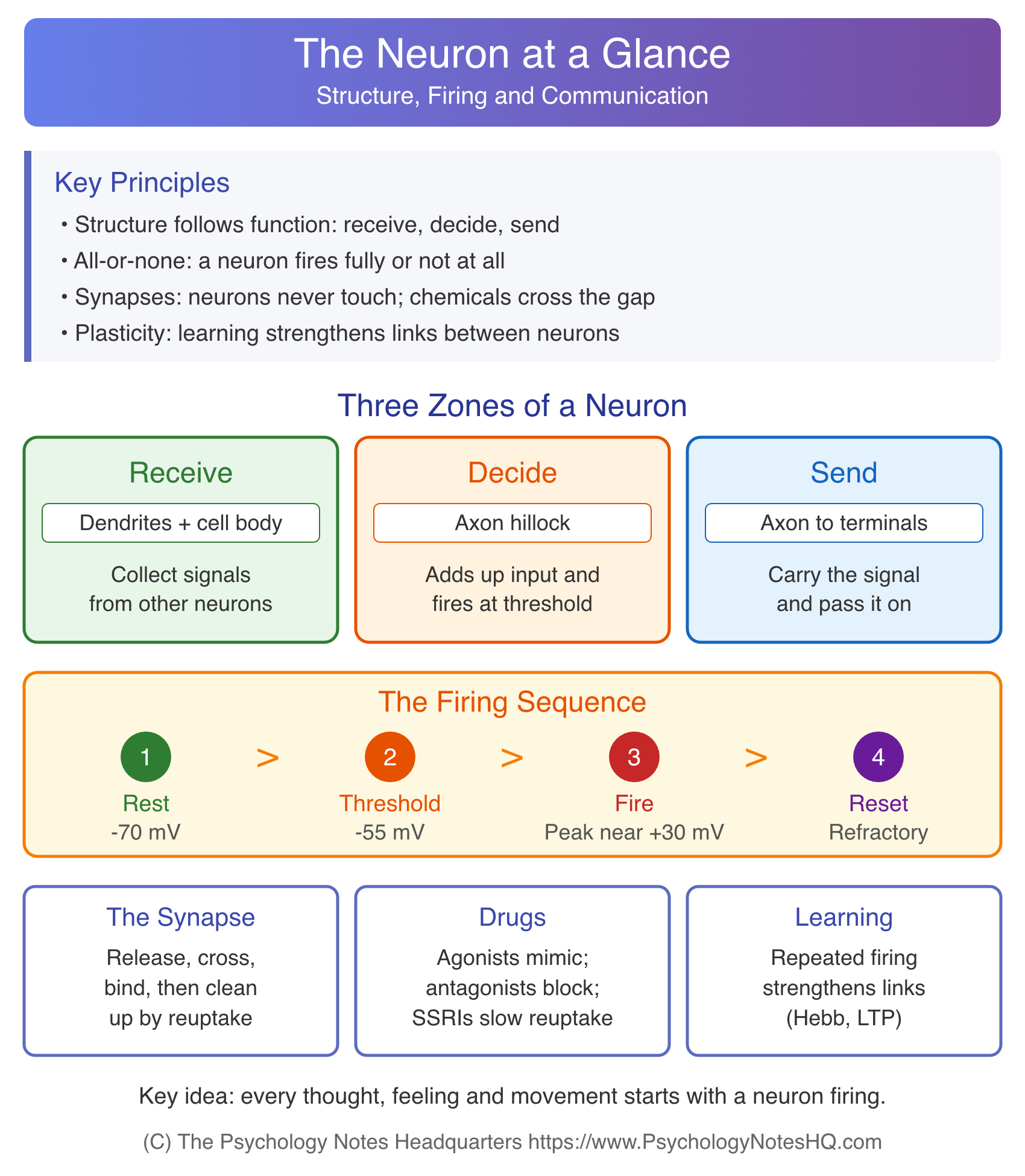
Every part of a neuron earns its place. Dendrites collect messages, the axon hillock decides whether to fire, and the axon, myelin and terminals deliver the result to the next cell.
Once you see the structure of a neuron as a system built for receiving, deciding and sending, the diagram stops being a list to memorize. It becomes a story you can explain, and that’s exactly what exam questions on biological psychology tend to reward.
References
- Azevedo, F. A. C., Carvalho, L. R. B., Grinberg, L. T., Farfel, J. M., Ferretti, R. E. L., Leite, R. E. P., Jacob Filho, W., Lent, R., & Herculano-Houzel, S. (2009). Equal numbers of neuronal and nonneuronal cells make the human brain an isometrically scaled-up primate brain. Journal of Comparative Neurology, 513(5), 532-541.
- Bliss, T. V. P., & Lømo, T. (1973). Long-lasting potentiation of synaptic transmission in the dentate area of the anaesthetized rabbit following stimulation of the perforant path. The Journal of Physiology, 232(2), 331-356.
- Fredholm, B. B., Bättig, K., Holmén, J., Nehlig, A., & Zvartau, E. E. (1999). Actions of caffeine in the brain with special reference to factors that contribute to its widespread use. Pharmacological Reviews, 51(1), 83-133.
- Hebb, D. O. (1949). The organization of behavior: A neuropsychological theory. Wiley.
- Hodgkin, A. L., & Huxley, A. F. (1952). A quantitative description of membrane current and its application to conduction and excitation in nerve. The Journal of Physiology, 117(4), 500-544.
- Purves, D., Augustine, G. J., Fitzpatrick, D., Katz, L. C., LaMantia, A.-S., McNamara, J. O., & Williams, S. M. (Eds.). (2001). Increased conduction velocity as a result of myelination. In Neuroscience (2nd ed.). Sinauer Associates.
- Spielman, R. M., Jenkins, W. J., & Lovett, M. D. (2020). Cells of the nervous system. In Psychology 2e. OpenStax.
How to cite this article:
The Psychology Notes Headquarters. (2026). Structure of a Neuron: 8 Parts, What Each Does, and the One Most Students Overlook. Retrieved from https://www.psychologynoteshq.com/neuron/